SHRI SHIVAJI SCIENCE COLLEGE, AMRAVATI
DBT STAR COLLEGE PROJECT ACTIVITY
ACTIVITY REPORT

Mounting of Sting Apparatus and Pollen basket of Honey Bee
Activity Dates: 24/09/2025
Type of Activity: Other
Organizing Department: Department of Zoology
Program Coordinators: Dr. G. A. Wagh; Dr. A. R. Rajoria
Head of the Department: Dr. J. D. Dhote
External Collaborator (if any): -
Objectives:
- 1. To isolate, mount and identify the anatomical structure.
- 2. To comprehend how the backward-facing barbs on the lancets operate in a sliding, reciprocating mechanism, enabling deeper stinger into the tissue.
- 3. To observe the fine structure of the lancet barbs under a microscope.
- 4. To understand the structural adaptations of the sting apparatus that ensures efficient venom delivery.
- 5. To demonstrate that the stinger is a modifies ovipositor, which is used for defense rather than egg- laying.
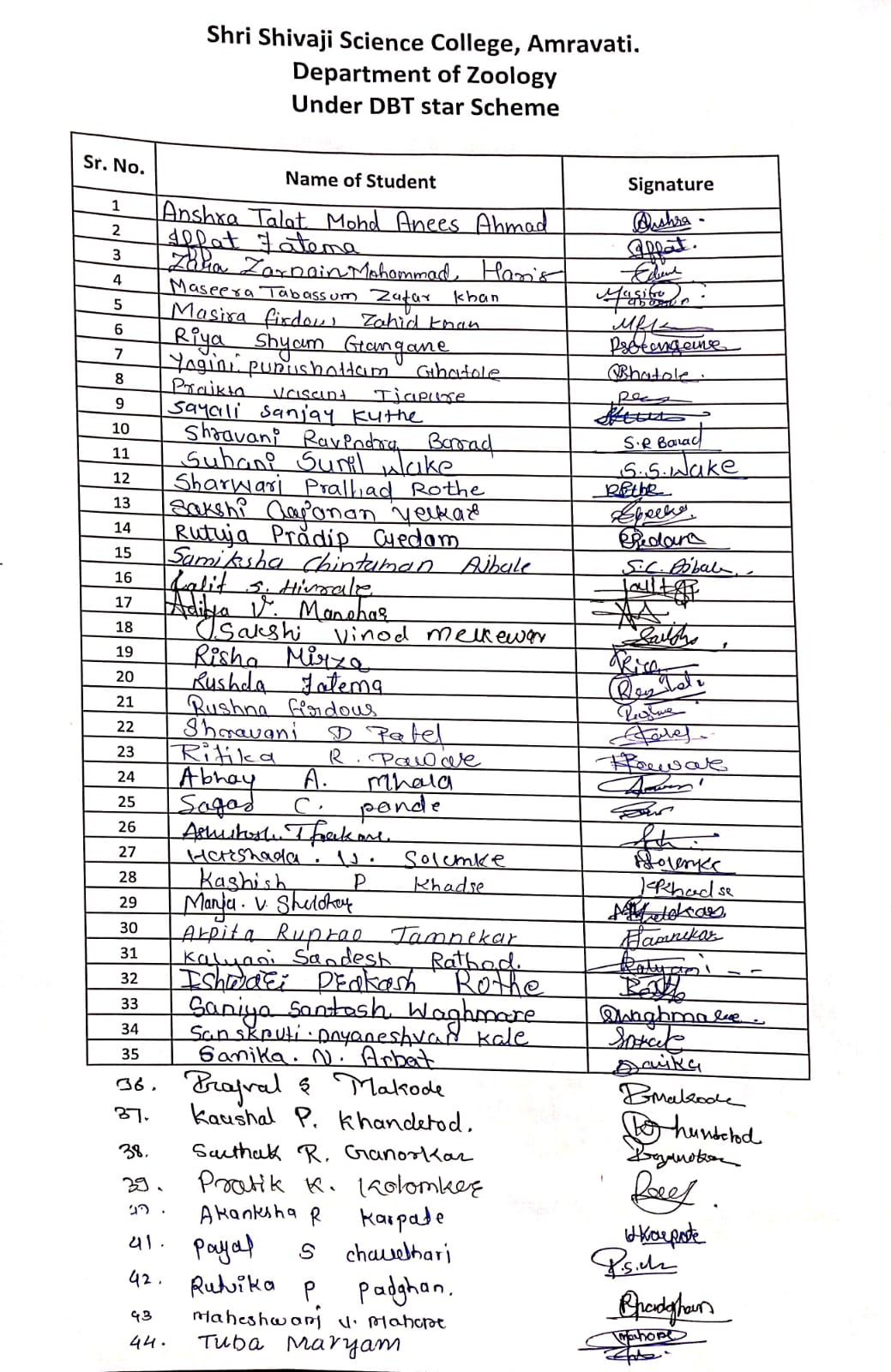
No of Beneficieries: 44
Classes Involved: Undergraduate Students
Venue of the Activity: Department of Zoology, Shri Shivaji Science College, Amravati
Activity Report:
To understand the importance of performing practical other than syllabus among students, Department of Zoology has designed and shortlisted some practical to be performed by Undergraduate students. This practice enables the students to widen their knowledge about their field. Before performing the practical, students were given the knowledge about the sting apparatus of honey bee including its structure, role etc.
Sting apparatus of honey-bee is modified ovipositor, found at the posterior extremity of abdomen in the worker bee. A honey bee’s sting apparatus includes a stinger with barbed lancet, a muscular bulb that pumps venom, a venom sac that stores it and a venom gland where it is produced. Located in the abdomen, the apparatus uses protractor muscles to drive the barbed stinger into the skin, while a valve within the bulb ensures venom is injected. The barbs prevent removal, causing the stinger and venom sac to tear from the bee’s body upon departure, leading to bee’s death. The bite of the sting causes burning sensation, pain and swelling of the part concerned. Preparation and mounting of honey bee sting apparatus was performed for UG students. For this, a detailed practical was chalked out and the sheets of the practical were distributed among the students for writing down. Students were already instructed to collect honey bee as well as already collected bees were used for the practical purpose.
After demonstration, students performed the practical by taking the provided worker honey bee and observed the posterior end of the abdomen under a dissecting microscope.
Students carefully, held the thorax with forceps and gently pull the last abdominal segment using a needle and extracted the sting apparatus without damaging it. This extracted sting apparatus was transferred to a clean slide with a drop of glycerin. The specimen was arranged properly with the help of a needle n the center of the slide and was mounted with DPX and a cover slip was gently placed over the specimen, avoiding air bubbles.
Detailed morphological and anatomical structures were observed by the students under a compound microscope and the outlined aims, objectives as well as outcomes were achieved.
This activity was successfully organized and conducted under the guidance of Dr. J. D. Dhote, Prof. & Head, Department of Zoology and Dr. G. A. Wagh, Prof. & Departmental DBT Coordinator, Dr. A. R. Rajoria, Asstt. Prof. & Activity Incharge, Dr. G. D. Hnde, Dr. S. V. Gawande, Dr. S. J. Kawade, Dr. P. M. Ramteke, Asstt. Prof. and non-teaching staff of department of Zoology.
Outcomes:
- 1. Students will isolate, mount and identify the anatomical structure.
- 2. Students will comprehend how the backward-facing barbs on the lancets operate in a sliding, reciprocating mechanism, enabling deeper stinger into the tissue.
- 3. Students will observe the fine structure of the lancet barbs under a microscope.
- 4. Students will understand the structural adaptations of the sting apparatus that ensures efficient venom delivery.
- 5. Students will understand the modification of the stinger as a modified ovipositor, which is used for defense rather than egg- laying.
Photos:
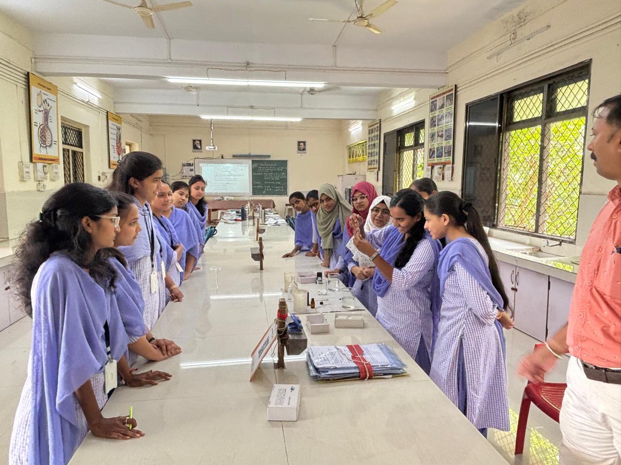 Students preparing for sting apparatus mounting of honey bee | 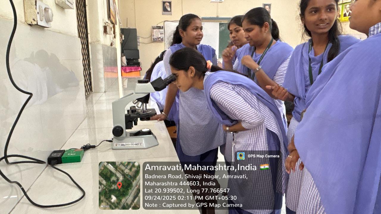 Students observing the slides |
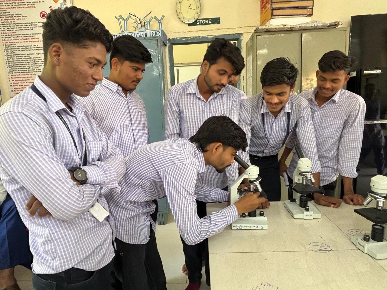 Students observing the slides under microscope. |  Students observing the mounted slides of sting apparatus of honey bee under microscope |
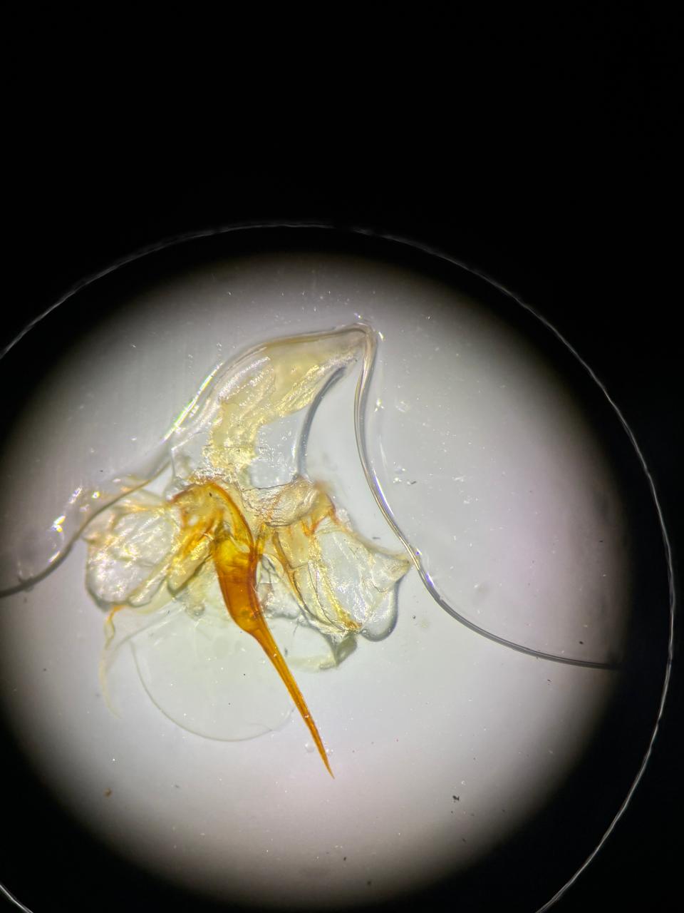 Mounted sting apparatus of honey bee seen in the microscope |  Instructions given to students regarding practical performance |
Attendance Sheet:
 Sheet |