SHRI SHIVAJI SCIENCE COLLEGE, AMRAVATI
DBT STAR COLLEGE PROJECT ACTIVITY
ACTIVITY REPORT
Field Visit Report: Department of Pathology & Blood Bank
Activity Dates: March 18, 2026
Type of Activity: Field Studies
Organizing Department: Department of Microbiology
Program Coordinators: Dr. Rasika R. Jane.
Head of the Department: Dr. R. C. Maggirwar
External Collaborator (if any): No
Objectives:
- To provide students with practical exposure to the functioning of a blood bank and pathology laboratory.
- To understand the procedures involved in blood collection, donor screening, and safe transfusion practices.
- To study the process of blood component separation and storage techniques
- To observe modern diagnostic techniques used in different sections of pathology such as histopathology, biochemistry, bacteriology, and mycology.
- To create awareness about the importance of voluntary blood donation and its role in healthcare.
No of Beneficieries: 50
Classes Involved: B.Sc.-II Year Microbiology students
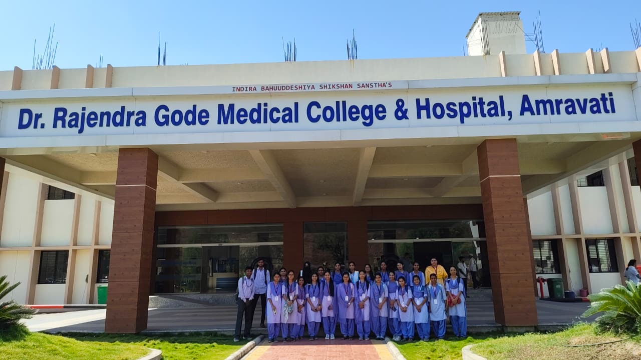
Venue of the Activity: Dr. Rajendra Gode Medical College, Hospital & Research Centre, Amravati
Activity Report:
Field Visit Report: Department of Pathology & Blood Bank
Institution: Dr. Rajendra Gode Medical College, Hospital & Research Centre, Amravati
Date of Visit: March 18, 2026
Participants: 40 Students (B.Sc. II Year)
Coordinator: Dr. Rasika R. Jane & Prof. Suwarna S. Mankar
1. Introduction
An educational visit to the Blood Bank and Pathology Laboratory was organized for students as part of their academic exposure to clinical diagnostic services and laboratory practices. The visit aimed to provide practical knowledge about blood collection, processing, testing procedures, storage techniques, and the role of pathology laboratories in disease diagnosis and patient care. Such visits help bridge the gap between theoretical learning and real-life laboratory functioning.
On March 12, 2026, the B.Sc. Second Year batch of 40 students undertook a specialized educational visit to the Dr. Rajendra Gode Medical College (RGMC), Amravati. This visit was organized to provide students with a practical understanding of the core clinical subjects—Pathology and Transfusion Medicine. Under the guidance of the college faculty, students explored the intricate workflows that bridge basic biological science with life-saving medical diagnostics.
The visit began with an orientation session in the blood bank, where students were introduced to the structure and working of a blood bank. The medical officer explained the importance of voluntary blood donation and how donated blood is safely collected, tested, processed, and stored for emergency use. Students observed the donor registration procedure, which includes recording medical history, checking haemoglobin level, blood pressure, pulse rate, and body temperature before blood donation. This screening ensures donor safety as well as recipient safety.
2. Comprehensive Analysis: The Blood Bank
The Blood Bank at RGMC serves as a critical lifeline for the hospital. The visit focused on the high standards of safety and precision required to manage human blood products.
A. Donor Selection and Phlebotomy
The process begins with Donor Screening. Students learned that not everyone can donate; a strict questionnaire regarding medical history, travel, and lifestyle is mandatory. Physical parameters such as Weight (>45kg), Hemoglobin (>12.5 g/dL), and Blood Pressure are recorded.
Phlebotomy: We observed the collection process using sterile, single-use blood bags. The most common anticoagulant used is CPDA-1 (Citrate Phosphate Dextrose Adenine), which maintains cell viability for up to 35 days.
B. Advanced Component Separation
Unlike smaller centers, RGMC utilizes a Refrigerated Centrifuge to separate whole blood into specific components. This "component therapy" allows one unit of blood to save up to three lives.
PRBC (Packed Red Blood Cells): Stored at 2°C to 6°C. Used for chronic anemia or acute blood loss.
FFP (Fresh Frozen Plasma): Stored at -30°C or colder. It contains clotting factors and is used for burn victims or liver disease.
Platelet Concentrate: Stored at 20°C to 24°C with constant agitation in a Platelet Agitator to prevent clumping.
C. Mandatory Screening and TTI Testing
Safety is paramount. Every unit is screened for Transfusion Transmitted Infections (TTI) using ELISA or CLIA methods. The mandatory tests include:
HIV I & II
Hepatitis B (HBsAg)
Hepatitis C (HCV)
Syphilis (VDRL)
Malaria (Smear/Antigen)
D. Immunohematology (Grouping & Cross-matching)
Students were shown the Gel Card Method for blood grouping, which is more accurate than the traditional slide method. We observed the Major Cross-match (Donor Cells + Recipient Serum) and Minor Cross-match (Donor Serum + Recipient Cells) to ensure 100% compatibility before the blood is issued to the wards.
3. The Pathology Laboratory: Departmental Breakdown
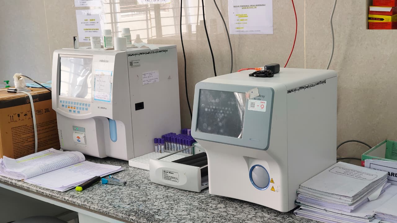
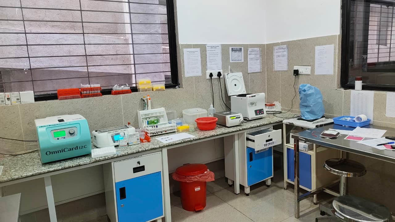
After visiting the blood bank, the students proceeded to the pathology laboratory. In the pathology section, they were introduced to routine diagnostic procedures such as blood testing, urine analysis, and microscopic examination. The laboratory staff explained the use of instruments like microscopes, centrifuges, automated analyzers, and haemoglobin meters. Students also learned about the importance of maintaining hygiene, sterilization, labeling, and proper sample handling to ensure accurate diagnostic results.
I. Histopathology: The Architecture of Disease
This department focuses on the microscopic study of diseased tissue.
Grossing: Pathologists examine large specimens (like an appendix or a tumor) and take "bits" for processing.
Tissue Processing: A 12-hour cycle involving dehydration (Alcohol), clearing (Xylene), and impregnation (Paraffin Wax).
Microtomy: We witnessed the use of a Rotary Microtome to cut sections as thin as 4 microns.
Staining: The H&E (Hematoxylin and Eosin) stain was demonstrated, where the Hematoxylin stains the nuclei blue/purple and Eosin stains the cytoplasm pink.
II. Biochemistry: The Chemical Blueprint
The Biochemistry lab is a hub of automation.
Analyzers: Students saw fully automated chemistry analyzers that perform hundreds of tests per hour.
Parameters: Key tests discussed included Serum Creatinine (Kidney function), Bilirubin (Liver function), and HbA1c (Long-term diabetes monitoring).
Quality Control: The staff explained the use of Levey-Jennings (L-J) Charts to monitor daily variations in machine accuracy.
III. Bacteriology: Identifying Microbial Threats
In the Bacteriology section, the focus is on isolating bacterial pathogens from clinical samples (blood, urine, pus).
Inoculation: We observed the streaking of samples on Blood Agar and MacConkey Agar.
Incubation: The use of aerobic and anaerobic incubators set at 37°C was explained.
Antibiotic Sensitivity (AST): Using the Kirby-Bauer Disc Diffusion Method, the lab determines which specific antibiotic will cure the patient's infection.
IV. Mycology: The Study of Fungi
With the rise in fungal infections, this department has become increasingly vital.
Identification: Students observed the characteristic fuzzy growth of Aspergillus and Candida on Sabouraud Dextrose Agar (SDA).
Microscopy: We viewed Lactophenol Cotton Blue (LPCB) mounts, which highlight the spores and hyphae of fungi under the microscope.
4. Conclusion and Educational Impact
The visit to Rajendra Gode Medical College provided an invaluable "hands-on" perspective that no textbook can replicate. The 40 students of B.Sc. II were able to correlate their syllabus with real-world medical practice. The transparency of the staff and the state-of-the-art infrastructure at RGMC offered a clear view of the career opportunities available in medical diagnostics and research.
Overall, the visit to the blood bank and pathology laboratory was highly informative and beneficial. It enhanced students’ understanding of diagnostic laboratory services and highlighted the importance of blood donation in saving human lives. The visit inspired students to appreciate the role of healthcare professionals working behind the scenes in patient diagnosis and treatment. Such educational visits are valuable in developing scientific awareness, practical knowledge, and social responsibility among students.
Outcomes:
- Explain the workflow and organization of a blood bank and pathology laboratory
- Describe the criteria for donor selection and the process of blood collection.
- Identify the major tests performed for detecting transfusion-transmitted infections.
- Recognize the functions of different pathology sections such as histopathology, biochemistry, bacteriology, and mycology.
- Develop awareness about career opportunities in medical laboratory sciences and healthcare.
Photos:
 Students during orientation session at the Blood Bank |  Observation of donor screening and registration process |
 Antibiotic Sensitivity Testing using Kirby-Bauer method | 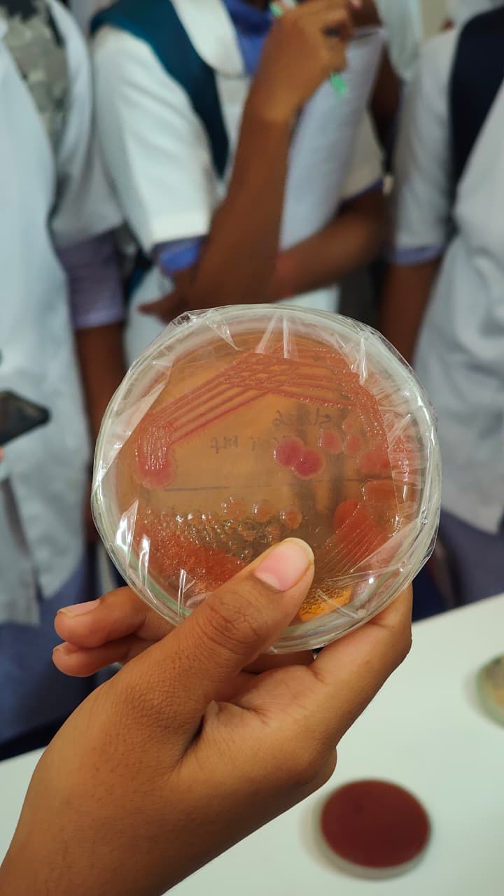 Group photograph of students at the Blood Bank and Pathology Department |
 Bacteriology lab showing culture plates (Blood Agar & MacConkey Agar) | 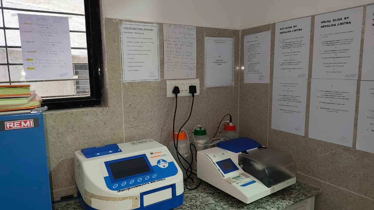 Fungal culture on Sabouraud Dextrose Agar (SDA) |
Attendance Sheet:
 |