SHRI SHIVAJI SCIENCE COLLEGE, AMRAVATI
DBT STAR COLLEGE PROJECT ACTIVITY
ACTIVITY REPORT
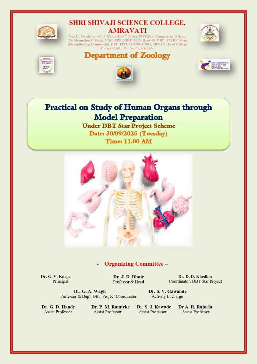
Practical Activity: Study of Human Organs through Model Preparation.
Activity Dates: 30th September 2025 (Tuesday)
Type of Activity: Practical Activity
Organizing Department: Department of Zoology
Program Coordinators: Dr. G. A. Wagh, Dr. S. V. Gawande
Head of the Department: Department of Zoology, Shri Shivaji Science College, Amravati
External Collaborator (if any): -
Objectives:
- To study the anatomical structure of major human organs.
- To develop 3D visualization skills through clay model preparation.
- To use creative educational tools (diagram cards and clay) to simplify complex biology topics
- To encourage group collaboration and scientific communication among students.
- To integrate practical, artistic, and theoretical skills in human anatomy learning.
No of Beneficieries: 41 Students
Classes Involved: B.Sc. III Year Students
Venue of the Activity: Department of Zoology, Shri Shivaji Science College, Amravati
Activity Report:
The Department of Zoology, Shri Shivaji Science College, Amravati, organized a practical activity titled “Study of Human Organs through Model Preparation” on 30th September 2025 (Tuesday) at 11:00 AM under the DBT Star College Scheme. The activity was conducted for B.Sc. III Year students with the guidance of Activity In-charge Dr. S. V. Gawande. The program was supported by the Head of the Department, Dr. J. D. Dhote, and coordinated under the Department DBT Coordinator by Dr. G. A. Wagh.
Understanding human anatomy is a fundamental requirement for students pursuing studies in life sciences. While theoretical knowledge forms the base, visual and practical exposure significantly enhances comprehension and retention. Keeping this in mind, the Department of Zoology organized a hands-on model preparation practical to promote experiential and interactive learning. The activity involved the preparation of three-dimensional models of human organs using dry super clay. To assist students, colourful diagram cards displaying labelled structures, color-coded parts, and brief functional descriptions were provided, enabling them to clearly visualize the organs before constructing their models.
The objectives of the practical were multifaceted. It aimed to help students understand the anatomical structure of major human organs in a detailed and engaging manner. The activity also focused on developing three-dimensional visualization skills through model preparation. By using creative educational tools such as diagram cards and clay, complex biological concepts were simplified. Furthermore, the activity encouraged group collaboration, communication, and the integration of theoretical knowledge with practical and artistic skills.
Adequate preparation and resources were arranged for the smooth execution of the activity. Materials such as multi-colours dry super clay, plastic boards, baseplates, markers, pens, glue, and printed diagram cards were provided to all participants. Students were divided into 16 pairs, making a total of 32 participants. Each group was assigned a specific human organ or system for model preparation. The selected organs included the human brain, heart, lungs, kidneys, liver, eye, ear, stomach, digestive system, and both male and female reproductive systems.
The practical session commenced with an introductory orientation by Dr. S. V. Gawande, who explained the objectives and demonstrated the use of diagram cards as reference tools. Students began by carefully studying the diagrams to understand the structure and functions of their assigned organs. They then proceeded to construct detailed 3D models using dry super clay, paying attention to proper size, proportion, and colour coding. To enhance clarity, labels were added using small tags to identify different parts such as atria, ventricles, optic nerve, cortex, and other anatomical features.
After completing the models, each group presented their work, explaining the structure, function, and significance of the organ they had created. This presentation not only reinforced their understanding but also helped in developing their communication skills. Faculty members evaluated the models based on anatomical accuracy, creativity, neatness, appropriate use of colour and labelling, and clarity of presentation.
The activity proved to be highly beneficial for the students. It enhanced visual learning by making internal structures easier to understand and remember. The hands-on experience allowed students to actively engage in the learning process, leading to better retention of knowledge. Working in pairs fostered teamwork, coordination, and collaborative learning. Additionally, the activity encouraged creativity, as students transformed theoretical concepts into tangible 3D representations, thereby bridging the gap between theory and practice.
The aims of the practical were successfully achieved. The activity enhanced students’ understanding of human organ structures, promoted visual and hands-on learning, and improved their ability to identify and label different parts accurately. It also encouraged creativity, teamwork, and scientific communication while supporting experiential learning aligned with the objectives of the DBT Star College Scheme.
In conclusion, the practical session was a successful and enriching learning experience. It highlighted the importance of innovative teaching methods in improving students’ understanding and interest in the subject. The enthusiastic participation and creative efforts of the students clearly reflected the success of the activity in achieving its educational goals and objectives.
Practical Activity:
Aim:
To study the structure and position of human organs by preparing models with the help of clay.
Requirements:
• Colour clay
• Cardboard or base sheet
• Pencil and eraser
• Adhesive (glue)
• Toothpicks or pins (for labelling)
• Sketch pens/markers
• Reference diagram of human organs
Procedure:
1. Select major human organs such as brain, heart, lungs, liver, stomach, and kidneys.
2. Take a cardboard or base sheet to prepare the model.
3. Prepare different organs using colored clay according to their shapes and sizes.
4. Arrange the organs on the base in their correct anatomical positions.
5. Fix them properly using glue.
6. Label each organ clearly using toothpicks or markers.
7. Compare the model with a reference diagram to ensure accuracy.
8. Study the structure and arrangement of the organs.
Observation:
• Each organ has a specific shape, size, and position in the body.
• The brain is located in the head region, the heart in the thoracic cavity, and lungs on both sides of the heart.
• The liver is on the right side, while the stomach is on the left side of the abdomen.
• The model provides a clear visual understanding of organ placement.
Result:
The clay model of human organs was successfully prepared, showing correct structure and arrangement of major organs.
Conclusion:
The preparation of a clay model helps in better understanding of human anatomy. It improves visualization, enhances learning, and makes the study of organs easy and effective.
Outcomes:
- To study the anatomical structure of major human organs.
- To develop 3D visualization skills through clay model preparation.
- To use creative educational tools (diagram cards and clay) to simplify complex biology topics.
- To encourage group collaboration and scientific communication among students.
- To integrate practical, artistic, and theoretical skills in human anatomy learning.
Photos:
 Models of human organs created by students | 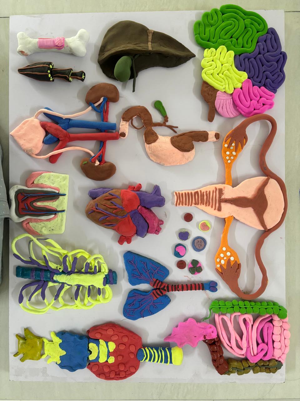 Students making clay models |
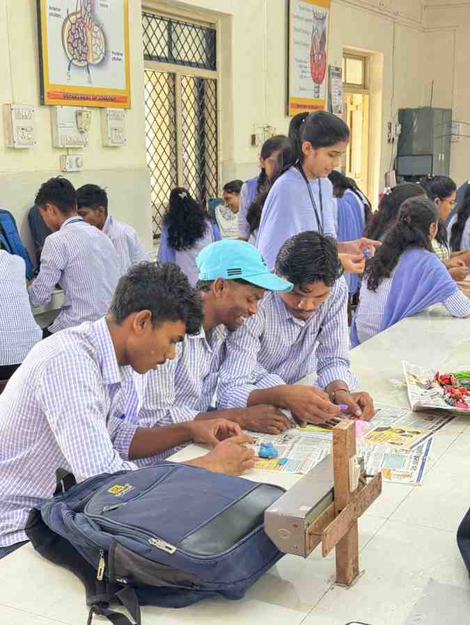 Students making clay models | 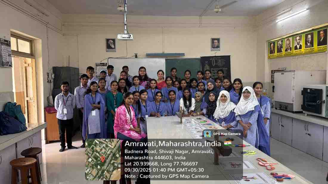 Group Photo of B.Sc. III Students |
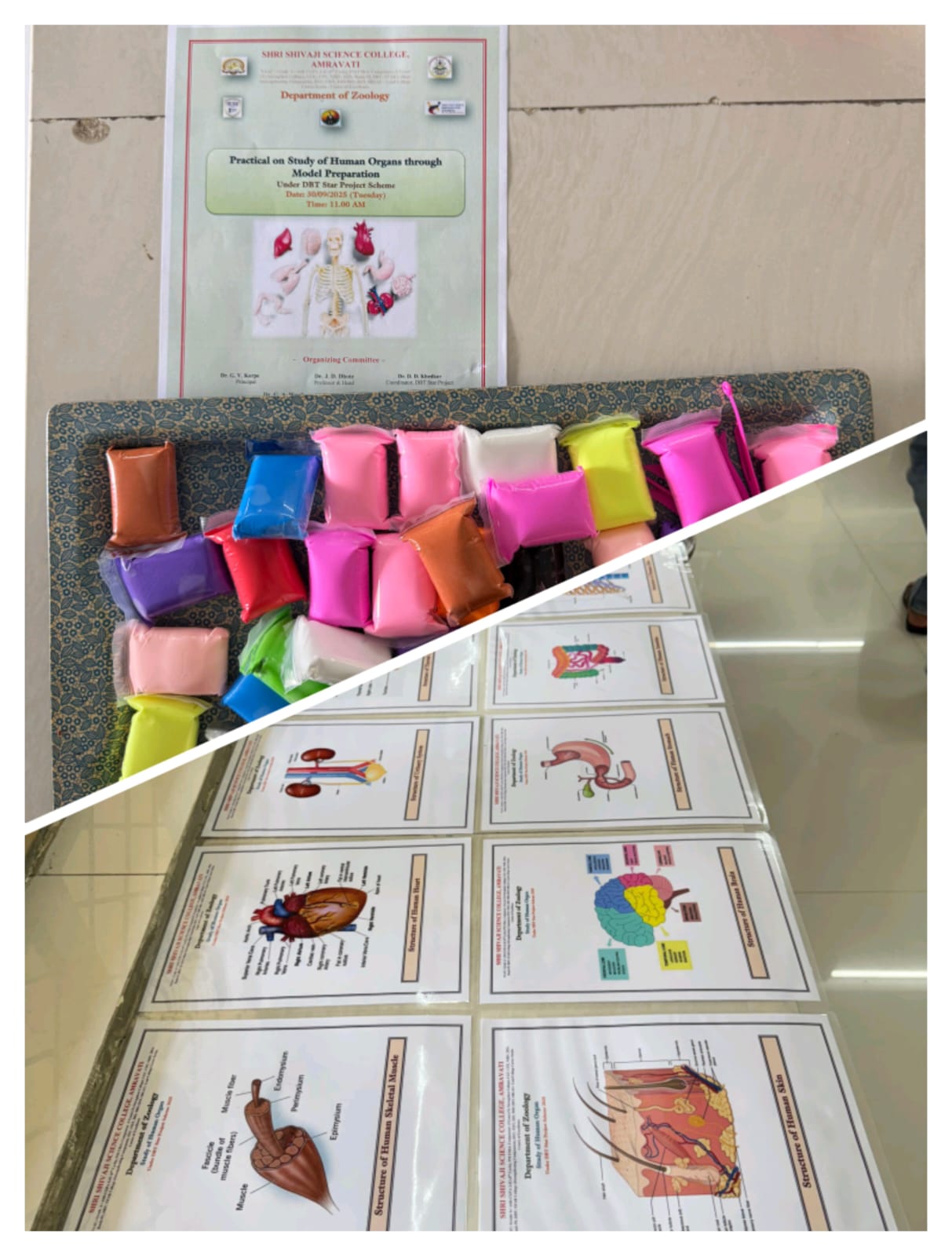 Materials used to create the model | 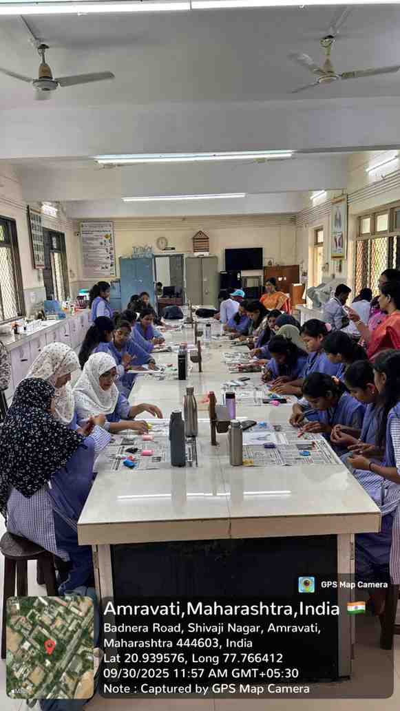 Students performing the first step of Clay Model |
Attendance Sheet:
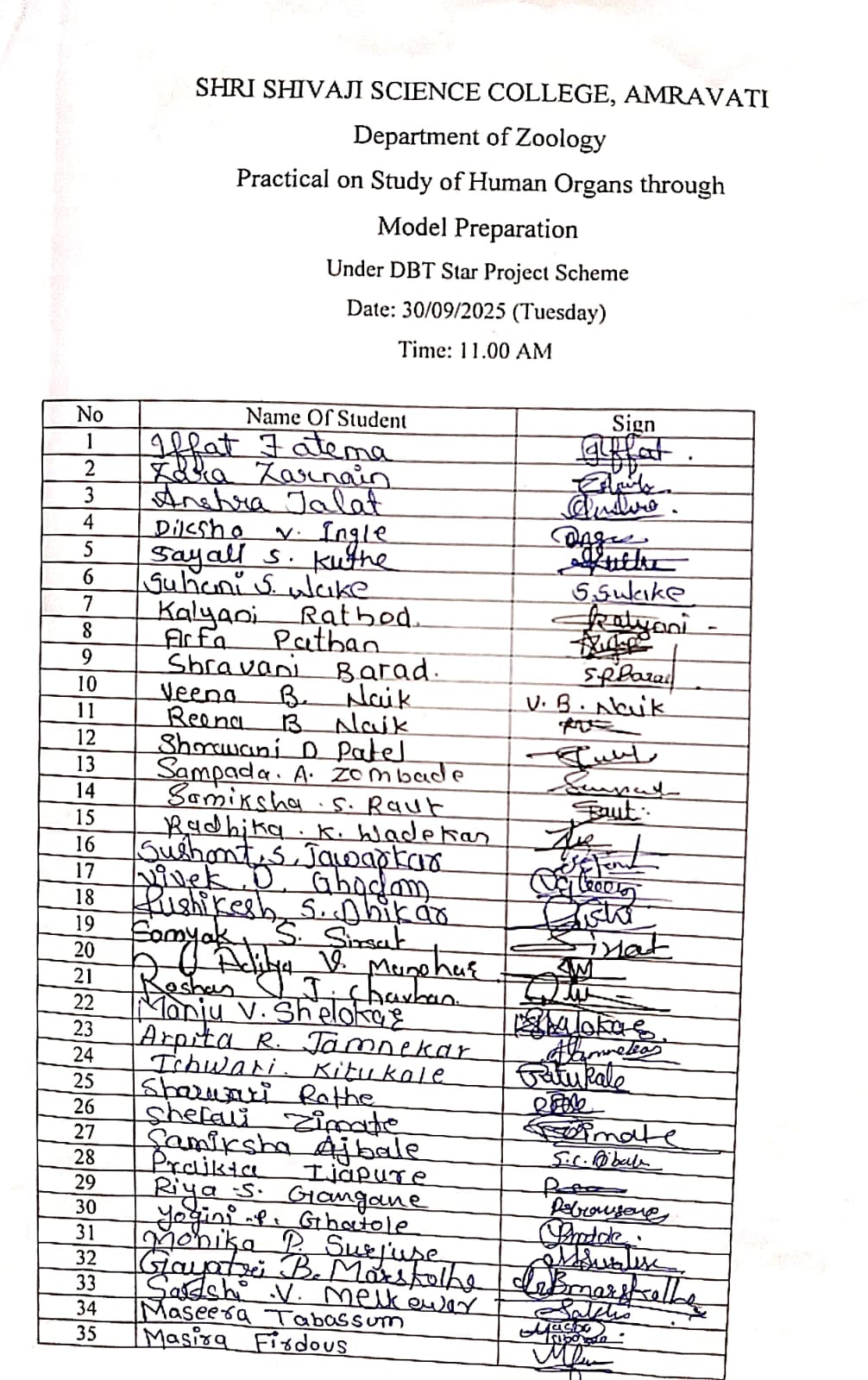 Sheet |
 Sheet |